
Radiology Normal Values Reference Guide: Ultrasound, CT & MRI
Normal measurements sit at the heart of every radiology report. Whether you are confirming a liver is within normal limits, deciding if a lymph node is pathologic, or checking that a pediatric kidney has grown appropriately for age, the difference between “normal” and “abnormal” often comes down to a number you need to recall in seconds.
This pillar guide pulls together the most frequently used normal reference values across ultrasound, CT, and MRI — for both adults and pediatric patients — into one reference you can bookmark. Each section below includes a quick-reference table, key clinical caveats, and links to deeper dives. Where values change with age, sex, or body habitus, we flag it rather than give a single misleading number.
Ultrasound Normal Values
Ultrasound is operator- and patient-dependent, so reference ranges are best treated as orientation rather than strict cutoffs. Measurements should always be made in a standardized plane, at end-inspiration where relevant, and compared against age- and body-size-adjusted norms in children.
Adult Abdominal Organs — Ultrasound Reference Ranges
| Organ | Measurement | Adult Normal Range |
|---|---|---|
| Liver | Craniocaudal span (midclavicular line) | Up to ~15–16 cm |
| Spleen | Long axis length | Up to ~12 cm (≤13 cm in tall adults) |
| Kidney | Bipolar length | 9–12 cm (each kidney; <2 cm side-to-side difference) |
| Pancreas | AP diameter (head / body / tail) | ≤3.0 / ≤2.5 / ≤2.0 cm |
| Common bile duct | AP diameter at porta hepatis | ≤6 mm (≤7–8 mm post-cholecystectomy; add 1 mm per decade >60) |
| Gallbladder | Wall thickness (fasting) | ≤3 mm |
| Abdominal aorta | Maximum AP diameter | <3 cm (≥3 cm = aneurysm) |
| IVC | Diameter at subcostal window | 1.5–2.5 cm with ≥50% inspiratory collapse |
| Portal vein | Diameter at porta hepatis | ≤13 mm |
| Thyroid lobe | AP diameter | ≤2 cm |
Obstetric & Pelvic Ultrasound Highlights
- Endometrial thickness — premenopausal: varies with cycle phase (≤4 mm menstrual to ~16 mm secretory); postmenopausal without bleeding: ≤8 mm; postmenopausal with bleeding: ≤4 mm.
- Ovarian volume — reproductive age: ≤~18–20 mL; postmenopausal: ≤~8 mL.
- Nuchal translucency — measured at 11w0d–13w6d; upper limit typically ≤3.0–3.5 mm depending on CRL.
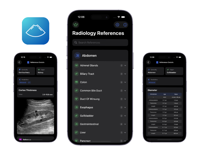
For the complete set of obstetric biometry (BPD, HC, AC, FL by gestational age), age-indexed pediatric organ sizes, and vascular Doppler cutoffs, the Radiology References app ships an offline, citation-backed database.
Try Radiology References Mobile App
Download our iPhone app for a better experience.

CT Normal Values
CT measurements are more reproducible than ultrasound but depend on phase of contrast, slice thickness, and reconstruction. Always measure in the axial plane perpendicular to the structure’s long axis unless otherwise specified.
Thoracic CT — Common Cutoffs
| Structure | Measurement | Upper Normal Limit |
|---|---|---|
| Thoracic aorta (ascending) | Maximum diameter | ≤4.0 cm (aneurysm ≥5.0 cm) |
| Thoracic aorta (descending) | Maximum diameter | ≤3.0 cm |
| Main pulmonary artery | Diameter at bifurcation | ≤2.9 cm (>2.9 cm suggests pulmonary hypertension) |
| PA:Aorta ratio | Axial | <1.0 |
| Mediastinal lymph nodes | Short-axis diameter | ≤10 mm (≤15 mm in subcarinal station) |
| Axillary lymph nodes | Short-axis diameter | ≤10 mm |
| Trachea | Coronal diameter (adult male/female) | ≤25 / ≤21 mm |
| Airway wall thickness | Segmental bronchi | ≤1.5 mm |
Abdominal & Pelvic CT
- Retroperitoneal lymph nodes — short-axis ≤10 mm; retrocrural ≤6 mm.
- Adrenal gland — limb thickness ≤5 mm; any nodule >1 cm warrants characterization.
- Appendix — outer diameter ≤6 mm; wall ≤3 mm.
- Small bowel wall — ≤3 mm (well-distended).
- Abdominal aorta — <3 cm; iliac arteries <1.5 cm.
- Pancreatic duct — ≤3 mm in head, ≤2 mm in tail.
Hounsfield Unit (HU) Quick Reference
| Tissue | Typical HU |
|---|---|
| Air | −1000 |
| Fat | −100 to −50 |
| Water | 0 |
| Simple cyst | 0 to 20 |
| Muscle / soft tissue | 30–50 |
| Acute blood | 50–80 |
| Adrenal adenoma (lipid-rich) | ≤10 (unenhanced) |
| Hepatic steatosis threshold | <40 HU (or liver >10 HU less than spleen on unenhanced) |
| Cortical bone | >700 |
MRI Normal Values
MRI reference values tend to be sequence- and field-strength-sensitive, but a core set of dimensional measurements recurs across almost every report.
Neuro MRI
- Lateral ventricles — Evans’ index (frontal horn width / maximal internal skull diameter) <0.3.
- Third ventricle — axial width ≤7 mm (up to 9 mm in the elderly).
- Fourth ventricle — AP diameter ≤12 mm.
- Pituitary gland — height ≤6 mm (prepubertal), ≤8 mm (adult male), ≤10 mm (adult female), up to ~12 mm in late pregnancy.
- Optic nerve sheath — diameter ≤5–6 mm (3 mm behind the globe); >6 mm suggests raised ICP.
- Corpus callosum thickness — genu ≥10 mm, splenium ≥8 mm in adults.
Spine MRI
- Cervical spinal canal AP diameter — ≥13 mm (≤10 mm = stenosis).
- Lumbar spinal canal AP diameter — ≥12 mm (≤10 mm = relative stenosis; ≤8 mm absolute).
- Conus medullaris tip — typically at L1–L2; below L2–L3 is considered low-lying in adults.
- Disc height — varies by level; asymmetric loss with endplate changes suggests degeneration.
Body & Pelvic MRI
- Prostate volume (PI-RADS v2.1) — calculated by ellipsoid formula; PSA density = PSA / volume, with >0.15 ng/mL/cc flagged as higher risk.
- Uterus — premenopausal up to ~8 × 5 × 4 cm; postmenopausal significantly smaller.
- Junctional zone of uterus — ≤12 mm on T2 (≥12 mm raises suspicion for adenomyosis).
- Rectal MRI (staging) — mesorectal fascia involvement defined by tumor within 1 mm of the fascia.
Pediatric Measurements: A Note on Age-Based Values
The single biggest trap in pediatric imaging is applying adult cutoffs to a child. Nearly every organ — kidney, spleen, liver, pancreas, thyroid, adrenal, ovary, uterus — has a normal size that scales with age, height, or body surface area. A 7 cm kidney is normal in a 2-year-old and suspicious in a teenager.
Key areas where age-indexed tables are essential:
- Kidney length — grows from ~4.5 cm at birth to ~10–11 cm by adolescence; nomograms exist for each month/year.
- Spleen length — approximately 6 cm at 3 months, 8 cm at age 3, 10 cm at age 10, with well-established upper limits.
- Liver span — by midclavicular line, scales roughly linearly with age.
- Adrenal gland — proportionally larger in neonates; limb thickness normative data differ from adults.
- Ovarian and uterine volumes — change dramatically through puberty; Tanner-staged normative data are the standard.
- Thymus — prominent and lobulated in infants; do not mistake for a mass.
Rather than memorize every curve, most pediatric radiologists keep age-indexed tables at hand. This is exactly what the Radiology References app was built for — pediatric values indexed by age group, with citations.
Try Radiology References Mobile App
Download our iPhone app for a better experience.
Using Normal Values in Daily Practice
A few practical tips that separate a defensible report from a shaky one:
- State the measurement technique. “Liver craniocaudal span at the midclavicular line: 17 cm” is reproducible; “liver enlarged” is not.
- Compare to patient-appropriate norms. Always ask: adult or pediatric? What body size? Is there a relevant comorbidity (e.g., cirrhosis shrinks the liver, chronic AF dilates the atria)?
- Report the cutoff, not just the number, when borderline. “Common bile duct 7 mm (upper limit of normal in this post-cholecystectomy patient)” gives the clinician the interpretation.
- Be explicit about uncertainty. Ultrasound-measured organ sizes can vary ±10–15% between operators. Repeat measurements and trend over time are more informative than a single absolute value.
- Cite your source when the value is contested. Useful for medicolegal defensibility and for trainees reading your report.
Carry This Reference in Your Pocket
This guide is intentionally broad a starting map of the territory. For the full set of measurements, organ-by-organ, modality-by-modality, indexed by age where it matters, with evidence citations and images, the Radiology References iOS app was designed to be the reference you reach for while reporting.