Easy Digital Bone Age Atlas for Children
Understanding bone age is crucial for pediatric healthcare who need to assess a child’s skeletal development and growth patterns. A bone age atlas serves as an essential diagnostic tool that helps determine whether a child’s bone development aligns with their chronological age, providing valuable insights into growth disorders, endocrine conditions, and overall health status.
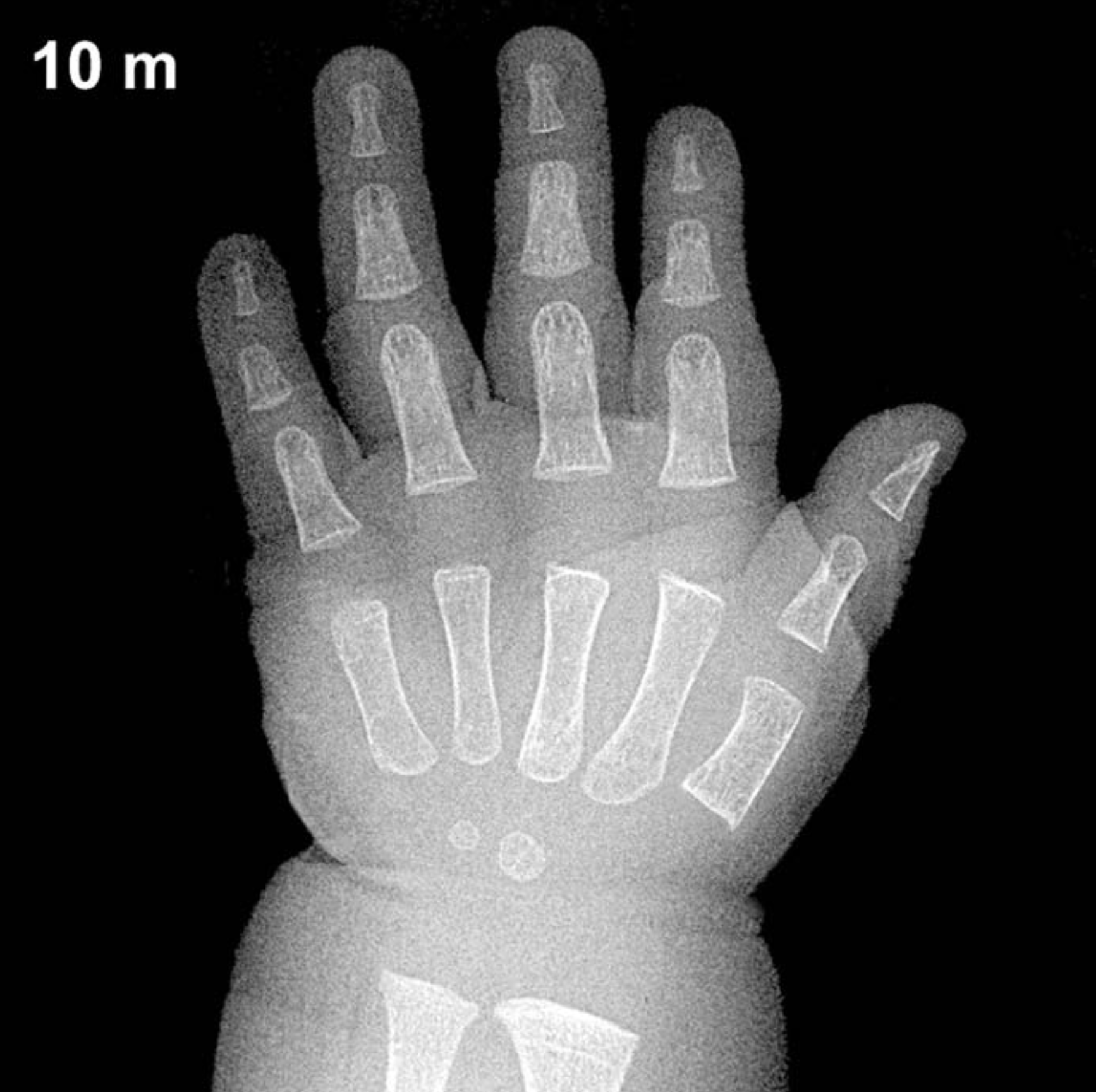
Traditional bone age assessment required extensive training, physical atlases, and significant time investment from healthcare providers. Our comprehensive digital bone age atlas revolutionizes this process by providing instant access to standardized reference images spanning from 8 months to 18 years for both male and female patients.
This interactive tool offers physicians an efficient way to compare patient X-rays against age-appropriate reference standards. With discrete age intervals matching actual developmental milestones, users can quickly identify the closest bone age match and make more accurate assessments.
Whether you’re a pediatrician evaluating growth concerns, an endocrinologist monitoring hormone therapy, or a medical student learning bone age assessment techniques, this digital atlas provides the comprehensive reference material you need for accurate, evidence-based evaluations.
How to Interpret Bone Age Atlas Images?
Key Structures to Examine
When analyzing bone age images, focus on three main areas of the hand and wrist. The carpal bones in the wrist are crucial indicators, as they appear and develop in a predictable sequence. The capitate and hamate bones appear first in infancy, followed by the triquetral, lunate, and scaphoid bones in early childhood. The pisiform bone is the last to develop, typically appearing around 8-12 years of age.
The metacarpal and phalangeal bones of the fingers provide important information about growth plate development. Look for the appearance and fusion of epiphyseal plates at the ends of these bones. The sesamoid bone at the thumb base is particularly significant, as it typically appears around 11-13 years and correlates with pubertal development.
The radius and ulna bones of the forearm offer additional assessment points. Examine their distal ends where they meet the wrist, paying attention to the shape and fusion status of the growth plates. These areas show distinct changes as children mature through adolescence.
What Changes to Look For
As children grow, bones undergo predictable transformations that help determine skeletal age. Initially, bones appear as small, round ossification centers that gradually enlarge and develop more defined shapes. The growth plates, visible as dark lines on X-rays, progressively narrow and eventually fuse completely when growth is finished.
Bone density and cortical thickness increase with age, making older bones appear whiter and more solid on X-rays. The overall proportions of bones also change, with epiphyses (bone ends) becoming more prominent and well-defined as children approach skeletal maturity.
Clinical Interpretation
Compare the patient’s bone development to age-matched reference images in the atlas. A bone age within 1-2 years of chronological age is typically considered normal. Significant advancement (more than 2 years ahead) may suggest precocious puberty or endocrine disorders, while significant delay (more than 2 years behind) could indicate growth hormone deficiency, hypothyroidism, or chronic illness.
Remember that girls typically develop faster than boys, with bone age often 1-2 years advanced compared to chronological age. This gender difference is normal and should be considered when making assessments. Always correlate bone age findings with the child’s clinical history, physical examination, and growth charts for the most accurate interpretation.
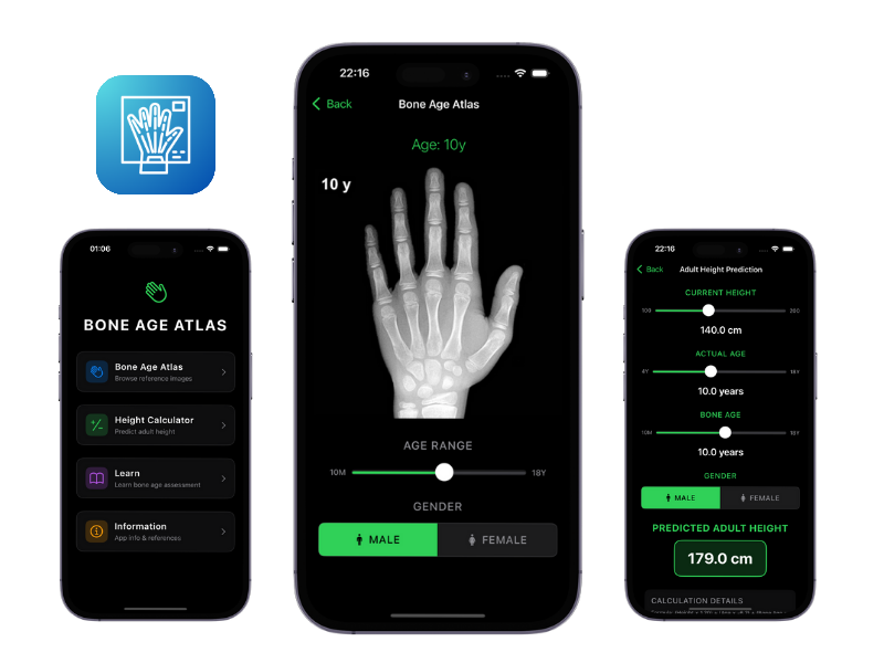
Try Easy Bone Age Atlas Mobile App
Download our iPhone app for a better experience.


Bone Age Atlas


